Sudden Painless Visual Loss

A sudden onset of visual loss is alarming to the patient. The diagnosis can be made in
most cases by a careful history and eye examination without immediate recourse to the
ophthalmologists, however, this may well be required. Only a few diagnosis require
immediate ophthalmic referrals for management:

  • central or branch retinal artery occlusion of less than 6 hours
  • any sudden visual loss of less than 6 hours and the cause can not be established
  • giant cell arteritis
  • All other causes of visual loss can be referred within 24 hours.
     
     
    History:
    • Transient visual loss like a 

    • curtain coming down (suggestive 
      of amourosis fugax)
       
    • Visual loss or field loss preceded

    • by sudden onset floaters and flashing 
      light (photopsia), this is suggestive of 
      retinal detachment
       
    • History of poorly controlled diabetes 

    • mellitus and laser treatment to the 
      retina (vitreous haemorrhage)
       
    • Headache +/- jaw claudication (pain 

    • in the jaw on eating) in the elderly 
      (giant cell arteritis)
       
    • Pain on eye movement in young patients 

    • (optic neuritis)
    Examination:
    • Visual acuity 
    • Assess the visual field by confrontation, 

    • some patients may have homonymous 
      hemianopia and yet complain of uniocular 
      visual loss.
       
    • Full ocular examination which should 

    • include:
       
      • pupil reaction for afferent pupillary

      • defect (this occurs in optic nerve
        disorder and extensive retinal
        pathology.)
         
      • retinal examination for any obvious 

      • signs
     
    The following are the most common causes of sudden visual loss seen in the casualty:


    Central or branch retinal artery occlusion

    Occlusion of the retinal artery may be caused by arteriosclerotic changes, embolus 
    (from heart or carotid artery) or inflammation (rare)

    History:

    • Sudden painless visual loss which may be complete (due to central retinal artery 

    • occlusion) or partial (due to branch retinal artery occlusion)
    • Patient usually have a history of hypertension or heart disease
    Examination:
    • The visual acuity is reduced in central retinal artery occlusion but may be normal 

    • in branch retinal artery occlusion
    • Relative afferent pupillary defect is present in central retinal artery occlusion
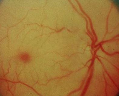
    • The retinal arteries are narrow or collapsed. 
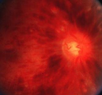
    • In central retinal artery occlusion, the fovea shows a cherry-red spot against the 

    • white infarcted retina. 
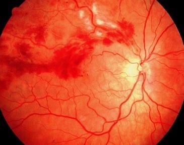
    • In branch retinal artery occlusion, the white infarcted retina corresponds to the 

    • occluded retina. 
    • Emboli may be seen in the arteries if the cause is emboli
    Management:
    • Immediate referral if the visual loss is less than 6 hours as treatment may restore 

    • some or most of the function. 
    • Treatment involves the use of intravenous acetazolamide and globe massage to 

    • lower the intraocular pressure and hopefully re-establish the arterial flow.
    • Further management aim to uncover any underlying diseases such as hypertension, 

    • cardiac or carotid thrombus. An ESR is usually performed in the absence of obvious 
      embolus to exclude arteritic causes.
    • Long term low dose aspirin is advised to reduce the risk of occurrence.
    Figure 1
    Central retinal artery occlusion showing the typical cherry-red spots again 
    the white infarcted retina. This appearance is not permanent and usually 
    disappears after about three days when the flow of the retina artery 
    is re-established but the retina does not regain its function.
    Figure 2
    Branch retinal artery occlusion due to emboli. Note their 
    presence in the inferior retinal artery. The infarcted 
    retina appears as white.

    Return to the top


    Central or branch retinal vein occlusion

    Retinal vein occlusion is a common vascular disorder caused by impaired venous blood flow. 
    It is second only to diabetes mellitus as a vascular cause of impaired vision.

    Presentation:

    • Sudden onset painless blurred vision
    • Less commonly the patient may present with painful red eye due to neovascular glaucoma
    • as a result of recent central retinal vein occlusion.
    Examination
    • The visual acuity is reduced in central retinal vein occlusion. However, the reduction is 
    • dependent on the severity of the occlusion. In branch retinal vein occlusion, the visual 

    • acuity may be normal if the fovea is not involved. 
    • Relative afferent pupillary defect may be present in patient with severe central retinal vein 

    • occlusion
    • Ophthalmoscopy reveals extensive intraretinal and pre-retinal haemorrhage with distended 

    • retinal veins.
    Management:
    • Refer within 24 hours.
    • Although there is no immediate treatment that can restore the vision, it is important to 

    • examine the patient for hypertension and glaucoma. A blood test is usually performed for 
      full blood count, ESR and in young patients auto-immune screening.
    • Follow-up in the clinic is arranged so that those at risk of neovascular glaucoma may be 

    • treated with laser pan-photocoagulation
    Figure 1
    A classical picture of central retinal vein occlusion showing extensive 
    flamed haemorrhages. 
    Figure 2
    This hypertensive patient complains of a sudden onset drop in his left vision. 
    Fundal examination reveals a left superior temporal branch retinal vein 
    occlusion. There are multiple flammed haemorrhages and cotton wool
    spots along  the occluded vein.
    Return to the page


    Retinal detachment

    History:

    • A recent history of floaters and flashes of light are common
    • Curtain coming across the vision 
    Examination:
    • Visual acuity variable depending if the macula is involved. If the vision is normal,

    • referral becomes more urgent as the macular function may be involved by the extension 
      of the detachment.
    • Visual field defect 
    • Ophthalmic examination in a dilated pupil shows greyish retina, hole and tear may be seen. 
    Management:
    • Refer the patient the same day 
    • Patients will require surgical management which consists of sealing the retinal breaks 

    • (using cryotherapy or laser) and relieving the vitreous traction (using indentation or vitrectomy)
    Figure 1
    This patient presented to the eye casualty with a sudden onset black shadow over his right 
    eye.Fundoscopy reveals a retinal detachment. Note the wrinkled retina; this is a common 
    sign of retinal detachment.
    Figure 2
    A patient with retinal detachment showing retinal tear.This patient requires treatment to seal
    the hole in order to prevent retinal detachment. This can either be achieved with laser or 
    cryotherapy. In patients who complain of sudden onset floaters, it can be difficult 
    to find abnormalities with a direct ophthalmoscope as the tears or holes are often in 
    the peripheral retina. Therefore, it is important to refer all patients with sudden onset floaters.


    Ischaemic optic neuropathy 

    In ischaemic optic neuropathy, there is occlusion of the small arteries around the optic disc. 
    It is important to differentiate arteritic optic neuropathy from non-arteritic optic neuropathy. 
    Arteritic optic neuropathy is caused by giant cell arteritis and prompt treatment with systemic 
    steroid can prevent involvement of the contralateral eye. 

    Presentation:

    • Sudden visual loss in a patient with a history of persistent headache (which may be 

    • temporal or occipital) or jaw claudication (pain in the jaw on eating) suggest giant cell arteritis
    Examination:
    • The visual loss is usually profound 6/60 or less in giant cell arteritis and less severe in 

    • non-arteritic ischaemic optic neuropathy.
    • Afferent pupillary defect is common
    • Fundal examination reveals swollen optic disc caused by occlusion of the arteries around 

    • the optic disc
    • In giant cell arteritis there is tenderness over the affected artery (usually the temporal artery) 

    • and the artery is usually not palpable.
    Management:
    • Refer immediately any patient with sudden visual loss and swollen disc for exclusion of 

    • giant cell arteritis.
    • ESR and the C reactive protein are usually raised in giant cell arteritis but non-specific. 

    • A definite diagnosis is by temporal artery biopsy for the typical granulomatous changes 
      in the arterial wall. However, systemic steroid is usually given while this is arranged.
         
    Figure 1
    Swollen optic disc with decreased vision and headache requires the exclusion of 
    giant cell arteritis. 


    Optic neuritis

    This condition typically affects patients in the 20 - 45 age group.

    Presentation:

    • Impaired vision 
    • Central field defect
    Examination:
    • Visual acuity may be as poor as perception of light
    • Central scotoma is typical
    • Impaired colour discrimination (best demonstrated with red object, the affected eye 

    • will see the red object less bright than the unaffected eye)
    • Relative afferent pupillary defect of the affected eye
    • Pain on eye movement especially on adduction
    • Fundal examination is normal as most cases have retrobulbar neuritis.
    Management:
    • Refer the patient within 24 hours for confirmation of the diagnosis
    • Normal or near-normal vision usually returns within 6 weeks
    • As treatment does not affect the outcome, unilateral optic neuritis is not treated by 

    • most ophthalmologists. However, follow-up is important as radiological investigation 
      may be needed to exclude compressive lesion in cases where spontaneous recovery 
      fails to recur.
       
    Figure 1
    Optic atrophy in a patient with a past history of optic neuritis. Optic atrophy is a late 
    sign. In acute optic neuritis, the optic disc is typically normal.
    Return to the top


    Vitreous haemorrhage 

    This may occur spontaneously or after trauma. The main causes of spontaneous vitreous 
    haemorrhage are: posterior vitreous detachment with or without retinal breaks, proliferative
    diabetic retinopathy, central retinal vein occlusion and subretinal neovascular membrane in 
    age-related macular degeneration with breakthrough bleeding.

    Presentation:

    • Sudden onset of floaters causing impaired vision
    Examination:
    • Depending on the severity of the haemorrhage, the view may be impossible with 

    • a direct ophthalmoscope
    Management:
    • Refer within 24 hours
    • In the casualty, the ophthalmologists will examine the eye for any posterior vitreous 

    • detachment and diabetic changes.
    • An ultrasound is often performed if the view is poor to exclude a retinal detachment
    • These patients require close observation until the cause has been established or treated
    Figure 1
    This diabetic patient complains of blurred right vision. Fundoscopy
    reveals a subhyaloid haemorrhage (haemorrhage between the retina
    and the vitreous). Note the haemorrhage has a fluid level (which is
    typical of subhyaloid haemorrhage) and the presence of previous laser 
    photocoagulation scars.This patient certainly has new vessels and 
    requires further laser photocoagulation.
     
    Return to the top