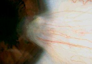
Pterygium is a degenerative condition associated with excessive
ultra-violet exposure. Clinically, it appears as a winged fibrovascular
tissue extending from the conjunctiva into the peripheral cornea.
Histologically, the conjunctival changes are indistinguishable
from that of pingueculae. The main differences between the two are mainly
clinical:
-
pinguecula can be present in both nasal and temporal bulbar conjunctiva
whereas pterygium is usually confined to the nasal aspect
-
pinguecula is confined to the conjunctiva whereas pterygium invade cornea
causing destruction of the Bowman's layer.
The following are the main features of pterygium:

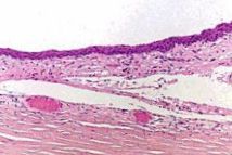
Normal conjunctiva with eosinophilic
stroma. |

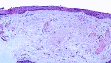
Pterygium with stromal elastosis.
(basophilic degeneration / staining) |
-
thinned conjunctival epithelium (occasionally this may be thickened)
-
elastotic degeneration of the stroma (the normal conjunctival stroma is
stained pink with H&E but in sun-damaged conjunctiva, the collagen
in the stroma is stained blue resembling the elastic tissue. This is also
called basophilic degeneration. This feature is also seen in the dermis
of actinic keratosis, basal cell carcinoma, squamous cell carcinoma and
melanoma)

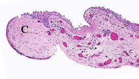
A specimen of pterygium containing corneal
stroma (C). The stroma of the conjunctiva shows
elastosis and presence of vasculature. |
Common viva questions:
-
What other lesions in and around eye may be associated with
sun damage?
-
What are the indications for removing pterygium? (Visual
disturbance, cosmesis and recurrent inflammation)
-
What measures may be used to reduce the recurrence rate of
pterygium? (conjunctival transplant to cover the bare area, beta radiation
and mitomycin)
|


