This is a common topic because of the easy availability of
corneal buttons taken from patients who underwent corneal transplant. As
most of these patients have severe keratoconus, the slides usually usually
show gross structural abnormalities.
In the slide look for:
-
disruption of the Bowman's layer
-
scarring in the stroma
-
thinning of the central stroma
-
rupture of the Descemet's membrane
-
presence of iron in the epithelium (Fleischer's ring)
-
absence of inflammation and vascularization
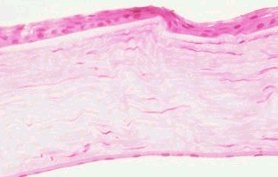
Low magnification H&E
Slides showing disruption of the Bowman's
membrane and stroma scarring. |
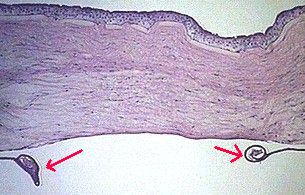
Low magnification
Stroma scarring with irregular corneal thickness and
rupture of the Descemet's membrane (Note the coiled
appearance, arrowed) |
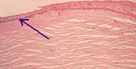
Low magnification H&E
Iron deposit in the epithelium (arrowed)
appearing dark blue. |
Common viva questions:
-
What conditions are associated with keratoconus?
-
What physical signs may be present in keratoconus?
-
How does hydrop develop and what is the treatment?
-
What do you know about the pathology of corneal graft rejection?
|