a
Optic nerve sheath meningioma
q
Like optic nerve glioma, you are more likely to be shown a gross specimen rather than a histological slide of optic nerve sheath meningioma. The most common topics for discussion are:
  • the differences between optic nerve meningioma and glioma
  • the presentation of optic nerve meningioma
The following are the main points about optic nerve meningioma:
  • orbital and optic nerve sheath meningioma are less common than intracranial meningioma. Orbital meningioma often arises secondarily from either intracranial meningioma or optic nerve sheath meningioma.
  • it is commoner in middle-aged female.
  • there is a small increased incidence in neurofibromatosis type I
  • clinical signs of optic nerve meningioma include unilateral papilloedema, optic atrophy and optico-ciliary shunt
  • there are a variety of  histological patterns but within the orbit only two are commonly found (see pictures below for description): 

  •  
    • meningotheliomatous
    • psammomatous

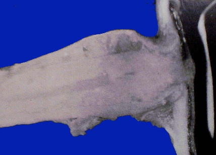
Unlike the fusiform appearance of an optic nerve glioma. The growth in optic 
nerve sheath meningioma tends to fill the subarachnoid space along the length
of the optic nerve. This give the tumour a railroad-track appearance on CT scan.


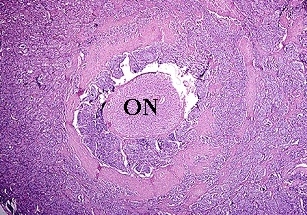
A cross section of optic nerve sheath meningioma
showing compression of the optic nerve (ON).


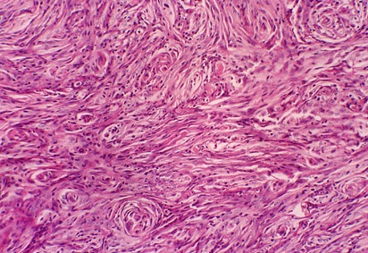
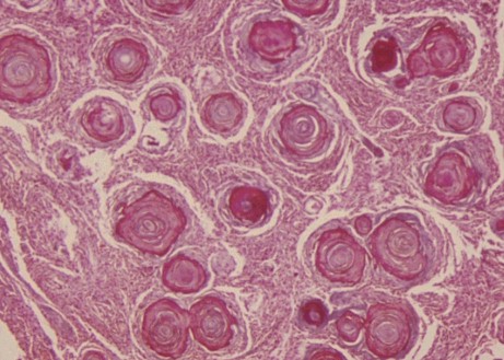
Meningotheliomatous type of meningioma. This is the most common cell type 
seen in all meningioma. The tumour has large, ill-defined cells with abundant 
cytoplasm. Numerous whorls are formed by several flattened cells wrapped 
around a large round cell. Psammomas bodies sometimes occur within the whorls.


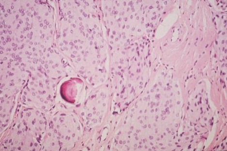
A single psammomas body within the meningioma. Psammomas body is made up 
of spherical calcified meningothelial cells.


This is a variant characterized by the abundance of psammoma bodies.These can
form confluent masses between which lie islands of meningothelial or fibroblastic
-like cells. This is seen less commonly than meningotheliomatous type.
 
Return to the main page