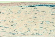
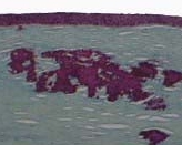
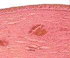
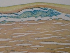
The above pictures are the typical appearance of the three
main stromal dystrophies with special stains. It is important that you
examine the actual slides under the microscope as the concentration of
the stains used and the microscopic lighting can alter the hue and saturation
of the colour.
The macular dystrophy can also be stained with colloid iron as seen
below.
If given a slide of lattice dystrophy, you are likely to be asked about
the phenomenon of birefringence and dichromism. Birefringence
occurs when the tissue stained with Congo red is viewed through a crossed
polarizing microscope. Dichromism occurs when the tissue is viewed with
polarized light and a green filter.
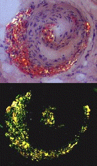
Birefringence means the amyloid can polarize (split) transmitted
light into two beams. Dichromism means two colour: the amyloid turns
from red to green depends on the orientation of the tissue under polarized
light. The picture below is a section of blood vessels with amyloid viewed
with and without polarized light.

