Plexiform neuroma
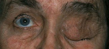 |
This patient has a left ptosis. The left upper is large
and containsa subcutaneous mass (classically described as felt like a bag
ofworms on palpation) which is immobile. (Note: in smaller plexiform neuroma,
the ptotic eye may assume a S-shaped deformity). This is
plexiform neuroma and is associated with neurofibromatosis
type I.
Look for other signs of neurofibromatosis:
-
there may be pulsatile exophthalmos due to absence of the
greater wing of sphenoid bone. The pulsation results from transmittedcranial
pulsation
-
skin lesions such as cafe au lait spots on the back and axiallary
freckles
-
afferent pupillary defect or ptoptosis from optic nerve glioma
-
on the slit-lamp look for Lisch's nodules
and ectopia uvea and examine the disc for cupping (the incidence of glaucoma
is high in patient with plexiform neuroma) and pallor (optic nerve glioma)
|
Questions:
1. How is type 1 neurofibromatosis transmitted?