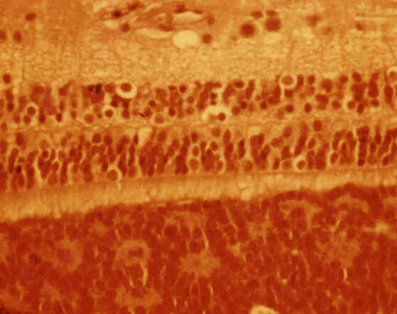
Figure 1 |

Figure 2 |
Paediatric Ophthalmology: Case two Special thank to Mr. Belgi for providing the pictures.

Figure 1 |

Figure 2 |
This one year baby was referred to the eye clinic because of suspected failure of visual development. Following fundoscopy an urgent CT scan was ordered.a. What does the CT scan show?
The CT scan shows bilateral solid masses within the globes with the density of the bone ie. calcification
b. What is the most likely diagnosis?In baby, the presence of calcified lesions within the globe should suggest retinoblastoma until proven otherwise.Retinoblastoma is the most common childhood intraocular tumour with an incidence of 1:20000. The disease is due to a mutation in chromosome 13q14. In 2/3 of cases the mutation is in the developing retina (somatic) and 1/3 in the germ cell (germinal). The former usually causes unilateral retinoblastoma where as the later bilateral multiple tumours.
c. What factors will determine the prognosis of this patient?Prognosis is determined by:
- optic nerve involvement. Involvement increases the incidence of metastasis to orbit, central nervous system and skull bones
- tumour size and location. Posteriorly located tumour tend to be detected earlier and therefore earlier treatment
- tumour differentiation. Presence of Flexner-Wintersteiner rosettes (Figure 2) suggest well-differentiated tumours and hence better prognosis
- age of the patient. Older patient tends to do badly due to delayed diagnosis
- secondary tumour. Bilateral retinoblastoma increases the chance of tumours like osteosarcoma and pinealoma
d. What is the chance of his offspring getting the same condition?The risk is about 40%.
Patients with bilateral retinoblastoma with or without familial history will transmit the disease to their offspring as an autosomal dominant trait with high penetrance (80%).
Click here for questions Click here for the main page Click here for FRCOphth/MRCOphth
/FRCS tutorials