The Ophthalmoscope
Why the pupil is black was a problem that attracted the attention of Roman writers, but the explanations they gave are merely of historical interest. It was held that the moisture in the eye was black; it was also suggested that the blackness resulted from the eye being a sort of deep trough. Yet the Ancients were also acquainted with the fact that some animal eyes are lustrous. Pliny observed that the eyes of nocturnal animals, such as cats, are brilliant in the darkness. The explanation had to wait for many centuries. Mariotte made some approach to it when he noted that the reason a dog's eye is luminous was that its "choroid" is white, and that hence the image of a light is painted on it clearly, whereas in man and in animals with black "choroid" no such clear image could be formed. This is a dim realization of the existence of the tapetum. Bidloo in the 17th century appreciated that no animal eye radiates light that it had not received, but it was not till 1810 that the simple observation that animal luminosity disappears in complete darkness was established by Prevost. This laid forever such views as those that regarded animal luminosity as a sort of phosphorescence; or that the radiation by night came from light absorbed during the day, or yet again that luminosity was the result of some such internal activity as is seen in the firefly. "Naked electricity" was yet another explanation that had passed muster. All these views had been invoked to explain the supposition that animals with lustrous eyes could see in the dark.Further advance towards a clearer understanding was supplied by the work of Rudolphi in 1821. He showed that the luminosity of an animal eye depended largely upon the direction of the ingoing rays. That furthermore the problem was a purely physical one he showed by the observation that the eye of a decapitated cat was just as effective for the production of luminosity as that of the living animal. A few years later Esser went further still by showing that the decapitated cat was really the better as the pupil was widely dilated.
That, at least partially, an optical problem underlay this animal luminosity was indeed also realized, more that a century earlier, when Mery in 1703 found that the luminosity of the cat's eye could easily be viewed when the animal was held under water. He appreciated that it was more than mere dilatation of the pupil consequent on suspended animation that was responsible for this phenomenon, and his explanation was that the water filled in unevenness of the cornea. The correct explanation was advanced by de la Hire six years later, when he argued that the cat's fundus was seen owing to abolition of corneal refraction under water; that consequently the rays emerged divergent, and some of them were thus caught by the observer's eye.
These considerations all seemed to have no practical significance. Even when luminosity in human eyes was observed the problem still remained an academic exercise. Duddell, in 1735, had noted the spontaneous luminosity of the eye of the human albino, as Woolhouse before him had observed it in white rats. Later in the century Fermin had noted luminosity of the eye of an Ethiopian albino (and incidentally held that this patient could thus see at night, because his eyes were like those of night animals). Further interest in spontaneous luminosity of man was aroused by Richter's observation (1790) that in one form of blindness luminosity was present. This led Beer to introduce the term amaurotic pseudo-glioma. Spontaneous luminosity was also noted in aniridia in 1829 by Beer.
No attempt to explain this spontaneous luminosity in man was made, though a close approach had been made to the explanation in the case of animals. The observation of Purkinje in 1823 that under certain conditions of illumination human eyes could be made luminous, passed unnoted. It had to be rediscovered independently by Cumming in 1846 and by Brucke in 1847. It was finally realized that the observer had to stand in the path of the emerging rays. Brucke indeed came near to inventing the ophthalmoscope when he looked through a tube placed in the flame of a candle illuminating the eye, and thus caught some of the emergent rays.
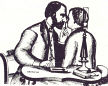
A conscious attempt to see the fundus was made by Kussmaul at about the same time (1845). On the basis of de la Hire's explanation of Mery's observation of the fundus of a cat submerged under water, he applied to the eye a plano-concave lens of the same power as the cornea, hoping to be thus enabled to see the optic nerve in the living human eye -- a procedure that " should be of great value in the diagnosis of certain eye diseases". He failed, for he did not realize the necessity for illuminating the eye. Babbage, of calculating machines fame, is another precursor; but whether he acted just as consciously as Kussmaul and what exactly he invented, is not definitely known. There is no documentary evidence as to what he made and what he showed to Wharton Jones in 1847, except the latter's account seven years later.
By this time the optical problem underlying luminosity of animal eyes and of the human eye under certain conditions had nearly reaches its solution. Indeed, the fact that the eye was not luminous under normal conditions because it forms an optical apparatus which returns entering rays to a focus at the source of illumination, had been indicated by the rather crude experiments of Kussmaul. THough he had failed to view the fundus in the living eye by neutralizing the refraction of the cornea, he showed that by further deranging the optical structure of the eye through removal of both the cornea and lens the fundus could be seen, and that it could likewise be seen if some vitreous was extracted and the retina came forward.
The crowning achievement came when Helmholtz announced the invention of an "eye-mirror" in December, 1850. His ophthalmoscope consisted not of a mirror but of plates of glass, four plates being used to increase the number of rays reflected into the eye. The illumination was of necessity poor. Modifications followed each other in rapid succession, the silvered mirror with a central hole arriving within a year. Two great improvements were likewise introduced at an early stage. Helmholtz's original ophthalmoscope was mounted with a holder for one lens, and lenses had to be changed constantly for eyes of different refraction. Rekoss, a technician, introduced a revolving disc carrying a series of lenses, whilst Ruete in 1852 introduced the indirect method of ophthalmoscopy. Thereafter an endless series of modification and improvements followed. The refracting ophthalmoscope was introduced at about 1870, whilst tentative electric ophthalmoscopes were brought out about fifteen years later, one of the earliest being that of Juler in 1886. Search for the ideal source of illumination led to attempts with oil, petrol, gas, daylight and almost every conceivable monochromatic flame.
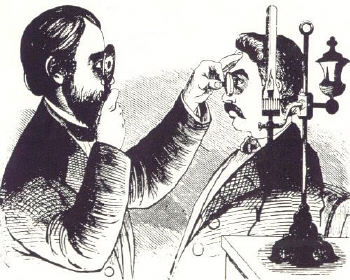
Early indirect ophthalmoscope. Note the inverted image is
illuminated with the light from a lamp placed on the table.The introduction of the ophthalmoscope in clinical ophthalmology was facilitated through it is brilliant application by von Graefe and his colleagues. Enthusiastically received by them it did not fare so well elsewhere. It was argued that it is dangerous for a diseased eye to be submitted to the strain of all this illumination. Some, more patronizingly, held that it might be quite a useful instrument for such oculists as have poor sight. Dixon of London, 1853, expressed the fear that its use might lead to amaurosis. In France, qualified support was given to the ophthalmoscope -- as indeed was the case with earlier models having a concave lens only. It was Anagnostakis who, in 1854, popularized the instrument in France by a series of excellent observations. In England, pioneer work was done by Spencer Watson, and ardent support came from Bowman, though as late as 1855 the Lancet could still speak sceptically of its value. In other countries, Holland excepted, it penetrated even more slowly. Yet by the time the First International Ophthalmological Conference was held in 1857 the ophthalmoscope had come to be sufficiently significant to claim the first discussion.

Ophthalmoscopes from the 19th century.Within a decade the ophthalmoscope had revolutionized ophthalmology. For one thing, it forced attention to the refractive state of the eye, supplying at the same time objective means of determining it. In no small measure the work of Donders is the result of the introduction of the ophthalmoscope. But even more far-reaching was the demolition of the age-long puzzle of amaurosis. At one stroke endless guesses, speculations, theories and discussions became meaningless. A new conception of glaucoma emerged early, even if at first it was held that in glaucoma no changes were present in the fundus, a view that was replaced by the belief that swelling of the disc was present, But by 1855 von Graefe, who with others had fallen into the earlier error as to swelling, demonstrated excavation and retinal pulsation - and iridectomy as a method of treatment of the hitherto hopeless and badly understood disease followed rapidly. A new chapter -- medical ophthalmology -- was opened by von Graefe in 1855 and Heymann in 1856 by the description of renal retinitis, whilst in 1860 von Graefe presented a boon to neurology by his observation of bilateral papilloedema. Coccius in 1853 described detachment of the retina and indicated retinitis pigmentosa. Thrombosis of the central vein was recognized by Liebriech in 1855, whilst von Graefe recognized embolism of the central artery in 1860.
...
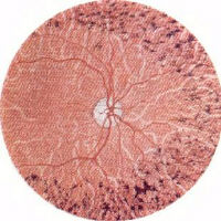
Early illustrations of the fundi from the 19th century when ophthalmoscopes
were first invented. The picture on the left shows advanced glaucoma and on
the right retinitis pigmentosa.Amaurosis, the condition which had been defined as one in which the patient saw nothing and the oculist also saw nothing , had ceased to exist.
| Return to the Main Page |