In pathology, granulomatous lesion is used to describe chronic inflammation
in which the predominant inflammatory cell is the macrophage with variable
amount of lymphocytes. The macrophages in such circumstances may aggregate
to form a circumscribed mass (granuloma). In addition, the macrophages
may fuse together to form multinucleate giant cells (different types of
inciting materials may produce different giant cells).
Granulomatous lesion typically occurs when the usual acute inflammatory
reaction involving neutrophils could not remove the inciting agent. The
second line of defense involving macrophages then take over. The common
inciting agents include the following:
-
Bacteria: mainly Mycobacteria such as TB or leprosy
-
Fungi
-
Exogenous foreign body
-
Endogenous altered material such as lipid (chalazion) or keratin (epidermoid/dermoid
cyst)
-
Unknown causes for example sarcoidosis
In the examination, the most common slides that feature granulomatous
lesions are:
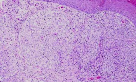
A skin specimen showing sarcoidosis. Note the presence
of giant cells. |
The granulomatous lesions in this disorder are typically non-caseating.
This is in constrast to TB granuloma which causes casating granuloma (slides
with TB granuloma is uncommon in MRCOphth).
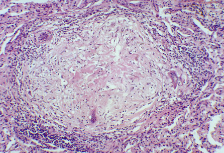
A TB granuloma showing central necrosis (caseating granuloma)
and the presence
of giant cells. |
|