Ocular pathology is an integral part of the final
MRCOphth / MRCS. However, most candidates probably have never seen a single
histopathology slide until a few months before the examination. It is recommended
that you should start examining the slides four to six months before the
examination to get use to using the microscope and analysing the slides.
Most teaching centre have a collection of ophthalmology slides for examination
and the pathologists are usually helpful and imaginative (for example who
else would have incorporated the following figure into their work? Click
on the figure for details.)
The examination usually takes the form of viva in which the candidates
are given specimen (but sometimes pictures) for examination and discussion.
The specimens may be a slide for microscopic examination (Figure 1) or
gross structural pathology(Figure 2) for naked eye examination.

Figure 1. Basal cell carcinoma |
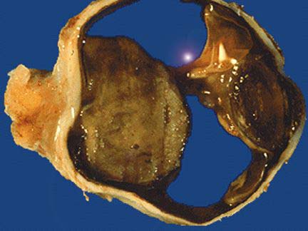
Figure 2. Choroidal melanoma. |
Although a wide range of specimens may appear in the examination, a survey
of candidates who sat the examination in the past five years shows some
clear favorite topics. In this section, only the commonly encountered specimens
are shown and discussed. If you like to contribute,
please e-mail to this address
chuaoxford@hotmail.com

Choroidal melanoma....
.

Retinoblastoma
|
a
a |
|