There is no substitute to examining pathology slides under
the microscope. Most teaching centres have histopathology slides for trainee
ophthalmologists. It is recommended that you examine the slides (especially
those mentioned in the main section) regularly before the examination to
understand the characteristic features for each condition.
When handed a slide, it is tempting to place it straight on the microscope
for detailed examination. However, in so doing some vital clues which are
visible to the naked eye may be missed.
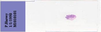
A piece of skin containing basal cell carcinoma. |
The following steps are recommended to derive the maximum information
from the slide:
-
Check the name and date of birth (sometimes the date the specimen was obtained
is written on the slide giving you clue to the patient's age)
-
Hold the slide against the light and decide on:
1. the type of tissue being examined (skin, cornea or the whole
eye. Avoid
guessing; ask the examiner for history if you were
not certain about its nature).
2. the type of stains used (this is especially useful in corneal dystrophy
as it
provides clue to the type of dystrophy being given).
3. the site and size of the lesion; intraocular tumour is always visible
on naked
eye (it is time-consuming to examine the entire
eye section under the
microscope, locating the site of the lesion
allows you to focus your
examination).
The history and the preliminary survey should have given you the diagnosis
or at least a differential diagnosis before you place the slide under the
microscope. The microscopic examination and findings will depend on the
pathology involved (details in relevant sections):
-
Skin tumours (decide if it is a benign or malignant tumour)
-
Other skin lesions (presence of giant cells or empty spaces as in chalazion
cells, fat cells in xanthelasma)
-
Choroidal melanoma (size and location, the cell types, any extraocular
invasion for example through the sclera or vascular invasion)
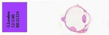
A naked eye examination shows the presence of a raised
intraocular
lesion and overlying retinal detachment (a choroidal
melanoma)
|
-
Retinoblastoma (size and location, differentiation, optic nerve invasion)
-
Cornea (bullae, pannus or intraepithelial basement membrane in the epithelium,
abnormal deposits, vessels, scarring or microbes in the stroma; break or
thickening of the Descemet's membrane; numbers of and pigmentation on the
endothelium)
|