This patient was referred by the optician because of this pigmented lesion. What features in the ultrasound favour a diagnosis of choroidal melanoma? Answer Elevated lesion with choroidal excavation on B-scan Low internal reflectivity (acoustic hollowness) on A-scan The ultrasound shows a lesion with a thickness of 1.9 mm and a base diameter of 7 mm with low internal reflectivity. What treatment options are there for this patient? Answer This is a small tumour and the treatment options include: Observation Photocoagulation Transpupillary thermotherapy Photodynamic therapy Plaque radiotherapy Proton-beam radiation Partial lamellar scleruvectomy Enucleation The patient refuses treatment and is managed by observation. What clinical features are predictive of growth of small choroidal melanoma? Answer The five main clinical features predictive of growth of small choroidal melanoma are: tumour thickness of greater than 2 mm posterior margin touching the optic disc visual symptoms orange pigment subretinal fluid The risk increases with the number of risk factors. (This is based on 1287 patients with small suspicious choroidal melanoma tumours measuring 3 mm or less in thickness, managed with observation. The follow up was 20 years with a median of 51 months. The results were published in Combination of clinical factors predictive of growth of small choroidal melanocystic tumours. Arch Ophthalmol 2000 Mar: 118 (3): 360-4 Shields CL et al.)
This patient was referred by the optician because of this pigmented lesion.
What features in the ultrasound favour a diagnosis of choroidal melanoma?
The ultrasound shows a lesion with a thickness of 1.9 mm and a base diameter of 7 mm with low internal reflectivity. What treatment options are there for this patient?
The patient refuses treatment and is managed by observation. What clinical features are predictive of growth of small choroidal melanoma?
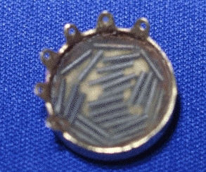
What is shown in the picture below? Answer Plaque containing radioactive rods used for plaque radiotherapy of small to medium sized choroidal melanoma. The plaque is sutured to the sclera over the lesion and leave for a few days before removing.
More questions