| Interpretation
of fluroescein angiography: I a |
 |
| When interpreting fluorescein angiography,
the simultaneous use of colour picture fundal is recommended. If you
were in a viva, ask the examiner if you could see the colour fundal picture.
In the OSE, you are likely to be given an enlarged single frame showing
characteristic pathology.
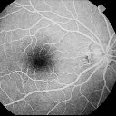
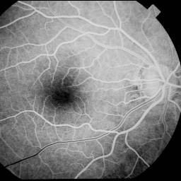
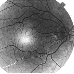
The fluorescein angiography may be given as a positive
or negative transparency (see picture below). As most fluorescein
angiographies encountered are on positive transparency, all the following
interpretations are based on the apparance on positive tranparency.
The fluorescein angiography is best viewed by placing the transparency on a viewing box and examine the frames with a 20 D lens (those used for indirect ophthalmoscopy). The frame also contains the time lapsed from the moment of intravenous fluorescein sodium injection. Fluorescein angiography can be analysed in the following ways (which complements each other):
it is examined frame by frames in the order that it was photographed. The major vascular phases of the angiogram is emphasized. This method is most useful in analysing vascular disorders of the retinal and choroidal. a observes each of the major layers of the posterior pose of the eye - the choroidal, RPE and neurosensory retina. a considers overall patterns. In an abnormal angiogram, some areas may be darker (hyperfluorescent) or lighter (hypofluorescent) than usually in a given location.
|
| Click for part II on interpration of FFA |
| Return to the main page |

